The x ray machine baggage integrates the principles of ergonomics with high-quality imaging technologies. The easy-to-use system enables users to navigate with ease. The system also integrates automatic calibration functionality to ensure accurate outcomes. The x ray machine baggage supports digital storage and retrieval solutions that offer healthcare providers easy access to diagnostic images.

The x ray machine baggage is used extensively in dentistry, yielding minute-level images of teeth, jaw bones, and surrounding tissues. It assists in the diagnosis of cavities, orthodontic problems, and impacted tooth diagnosis. The x ray machine baggage is used for endodontic and implant planning to allow precision treatment delivery.

The x ray machine baggage of the future will target integrating artificial intelligence to aid image interpretation and identify anomalies. Analysis software will automatically detect early-stage diseases more accurately. The x ray machine baggage will further feature low-dose radiation technologies, which will ensure that imaging is safer, more sustainable for both patients and operators.
The x ray machine baggage needs regular maintenance to function at its best. Technicians need to regularly inspect exposure controls, cooling systems, and image sensors. The x ray machine baggage has to be run within prescribed usage boundaries, and annual recalibration needs to be planned to maintain radiation accuracy as well as uniform imaging quality.
The x ray machine baggage has been used heavily in various medical fields due to its ability to offer rapid and precise medical images. The x ray machine baggage offers precise images of the various body parts that help in the diagnoses of various conditions such as bone injuries, cancer, and infections. The x ray machine baggage uses advanced imaging softwares that offer high contrast images.
Q: What makes an x-ray machine different from a CT scanner? A: An x-ray machine captures a single 2D image, while a CT scanner takes multiple x-rays from different angles to create 3D cross-sectional views. Q: How is image quality measured in an x-ray machine? A: Image quality depends on factors like contrast, resolution, and exposure settings, which are adjusted based on the target area being examined. Q: What power supply does an x-ray machine require? A: Most x-ray machines operate on high-voltage power systems, typically between 40 to 150 kilovolts, depending on their intended use. Q: Can x-ray machines be used for dental imaging? A: Yes, specialized dental x-ray machines provide detailed images of teeth, jaws, and surrounding structures to support oral health assessments. Q: How does digital imaging improve x-ray efficiency? A: Digital systems allow instant image preview, faster diagnosis, and reduced need for retakes, improving workflow efficiency in clinical environments.
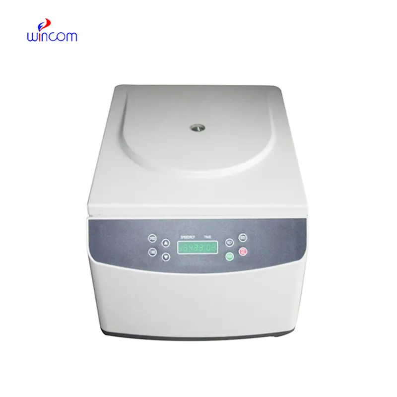
We’ve used this centrifuge for several months now, and it has performed consistently well. The speed control and balance are excellent.
This x-ray machine is reliable and easy to operate. Our technicians appreciate how quickly it processes scans, saving valuable time during busy patient hours.
To protect the privacy of our buyers, only public service email domains like Gmail, Yahoo, and MSN will be displayed. Additionally, only a limited portion of the inquiry content will be shown.
Could you please provide more information about your microscope range? I’d like to know the magnif...
We’re currently sourcing an ultrasound scanner for hospital use. Please send product specification...
E-mail: [email protected]
Tel: +86-731-84176622
+86-731-84136655
Address: Rm.1507,Xinsancheng Plaza. No.58, Renmin Road(E),Changsha,Hunan,China

 af
af
 es
es
 ar
ar
 tr
tr
 sw
sw
 pt
pt
 th
th
 ur
ur
 bn
bn
 ne
ne
 vi
vi
 km
km
 lo
lo
 de
de
 ru
ru
 fi
fi
 nl
nl
 fa
fa
 fr
fr
 ko
ko