With video displayed through beamforming and noise-filtering technology, the wireless probe ultrasound scanner is able to present an image that is very sharp and stable. Easy-to-use touch-screen controls help to streamline the process while rapid image rendering is guaranteed by the fast processing. Equipped for contemporary healthcare settings, the wireless probe ultrasound scanner is capable of working with both 2D and Doppler imaging.
The vast clinical applications of the wireless probe ultrasound scanner technology made it possible for nephrology to monitor kidney function efficiently and detect abnormalities in kidney structure. In the frontiers of endocrinology, the obtained data can reveal even the smallest nodules in the glands. The wireless probe ultrasound scanner is also a surgical device for blood flow patterns and vessel integrity.
The wireless probe ultrasound scanner should integrate with intelligent diagnostic ecosystems and communicate effortlessly with smartphones and electronic records. The synchronized exchange of data in real-time should enable constant patient observation. The next version should focus on improved design, better processing power of artificial intelligence algorithms, and enhanced reconstruction functions.
For long-term functionality, it is recommended that the wireless probe ultrasound scanner remain within an environment that maintains controlled levels of both humidity and temperature. The cables should be unwound slowly to ensure that no undue stress or wire breakages occur. The wireless probe ultrasound scanner should also be properly disinfected each time a patient has been examined.
Designed to be accurate and functional, the wireless probe ultrasound scanner offers high-definition imaging for diagnosis in numerous medical settings. It is comfortable with obstetric, vascular, and abdominal procedures and delivers exceptional definition. The wireless probe ultrasound scanner increases the certainty of diagnosis and reduces patient disruption through its non-invasive mode of operation. Its digital components allow for storage of data, transfer of images, and analysis.
Q: What makes the ultrasound scannert effective for diagnostic imaging? A: Its high-frequency sound wave technology allows accurate visualization of internal body structures in real time. Q: How portable is the ultrasound scannert? A: The device features a compact and lightweight design, allowing easy movement between clinical departments. Q: What types of probes are compatible with the ultrasound scannert? A: It supports multiple probe types, including linear, convex, and phased array probes for varied diagnostic needs. Q: Does the ultrasound scannert require special training to operate? A: Basic technical training is recommended to maximize its imaging performance and functionality. Q: How long can the ultrasound scannert operate continuously? A: It is designed for extended use with efficient cooling systems and stable power performance.
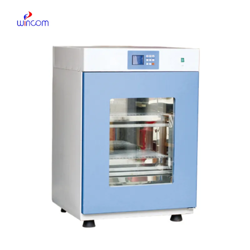
The centrifuge operates quietly and efficiently. It’s compact but surprisingly powerful, making it perfect for daily lab use.
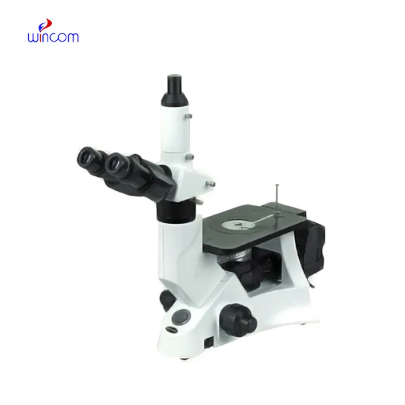
I’ve used several microscopes before, but this one stands out for its sturdy design and smooth magnification control.
To protect the privacy of our buyers, only public service email domains like Gmail, Yahoo, and MSN will be displayed. Additionally, only a limited portion of the inquiry content will be shown.
We’re interested in your delivery bed for our maternity department. Please send detailed specifica...
We’re currently sourcing an ultrasound scanner for hospital use. Please send product specification...
E-mail: [email protected]
Tel: +86-731-84176622
+86-731-84136655
Address: Rm.1507,Xinsancheng Plaza. No.58, Renmin Road(E),Changsha,Hunan,China

 af
af
 es
es
 ar
ar
 tr
tr
 sw
sw
 pt
pt
 th
th
 ur
ur
 bn
bn
 ne
ne
 vi
vi
 km
km
 lo
lo
 de
de
 ru
ru
 fi
fi
 nl
nl
 fa
fa
 fr
fr
 ko
ko