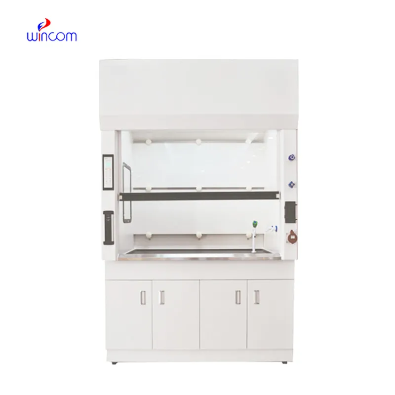
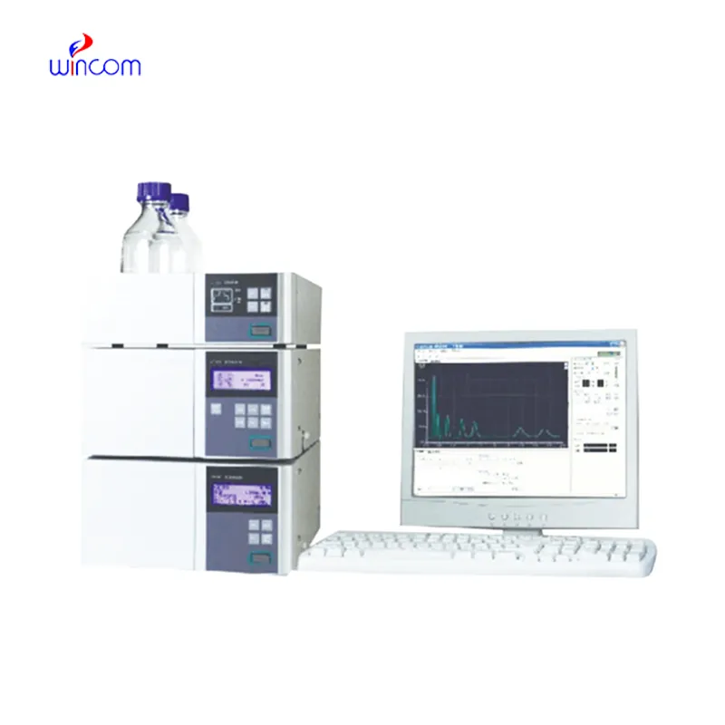
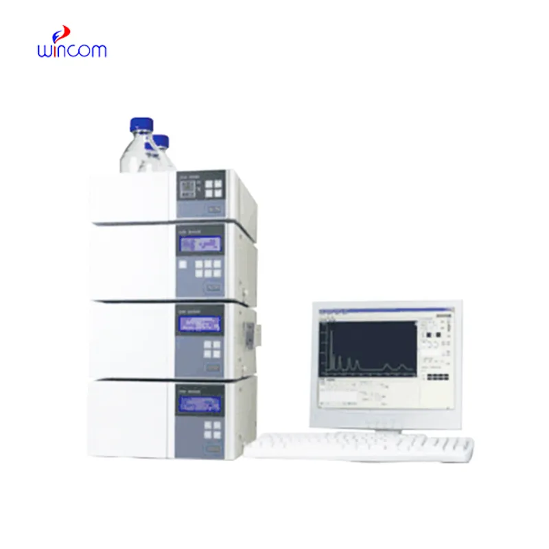
rp-hplc is a critical technique to obtain analytical information in studies of medicines, clinical samples, and biochemistry. It isolates compounds according to their chemical characteristics, generating reproducible analytical results. Laboratory scientists use rp-hplc to perform drug stability tests, monitor patient biomarkers, and find impurities. Its very high accuracy and flexibility allow thorough sample analysis in research, hospital, and clinical laboratory environments, thus becoming a fundamental device for assuring precision in both experimental and diagnostic results.
rp-hplc finds extensive application in hospital laboratories for monitoring drugs therapeutically. It provides precise determination of drug levels in patients' samples, thus making safe and effective dosing possible. Metabolites are tracked, treatment progress is assessed, and unexpected drug interactions are detected by the laboratory personnel. Its high accuracy and reproducibility facilitate both medical decision-making and research, hence, rp-hplc becomes an indispensable instrument in taking care of patients and analyzing the medical field.
rp-hplc is expected to have an increasing role in personalized medicine, analyzing complicated biomarkers swiftly. In the future, their application in hospitals will be centered on integrating pharmacokinetics, metabolomics, and monitoring, helping medical practitioners have access to swift and comprehensive data. The workflow in laboratories is expected to be organized.
The effectiveness of a laboratory is determined by the proper maintenance of rp-hplc. If the pump seals are regularly cleaned, the flow rates are monitored, and the usage of incompatible solvents is avoided then damage to the laboratory equipment can be prevented. It is essential for the technicians to carefully examine the columns, detectors, and tubing and in case of any sign of wear to conduct the scheduled calibration. Keeping rp-hplc in their best condition guarantees reproducibility, lowers the risk of equipment breakdown, and provides continuous performance for both hospital tests and experiments.
rp-hplc are a major factor in the daily activities of pharmaceutical labs, as they are used for verifying drug formulations, detecting impurities, and making sure that quality standards are met. It provides accurate quantification by separating active ingredients from excipients. Lab scientists utilize this for process optimization and stability evaluation under varied conditions. By providing reproducible analytical data, rp-hplc assists in both method validation and research development. Its accuracy guarantees that pharmaceutical products will be compliant with regulations. In lab environments, rp-hplc is a time-saving method not only for compound profiling but also for comprehensive analyses, thus being a fundamental tool in the quality control of pharma and research labs dealing with drug development.
Q: Do you need special training for HPLC operation? A: The answer is yes, training is a prerequisite to accurately and safely using pumps, columns, and detectors. Q: What type of maintenance does HPLC have? A: It requires cleaning, flushing, and inspection of all components as well as calibrating. Q: Is it possible to use HPLC in drug monitoring? A: Sure, it is a common practice in hospitals to monitor the levels of therapeutic drugs and also to identify metabolites in the samples taken from the patients. Q: What is the duration of analysis using HPLC in a typical case? A: The analysis time can range from a few minutes to more than an hour depending on the nature of the sample and the kind of column used. Q: Is HPLC a good choice for environmental testing? A: Yes, it can be used to find out the presence of pollutants, pesticides, and other harmful substances in water, soil, and air samples.
The water bath performs consistently and maintains a stable temperature even during long experiments. It’s reliable and easy to operate.
I’ve used several microscopes before, but this one stands out for its sturdy design and smooth magnification control.
To protect the privacy of our buyers, only public service email domains like Gmail, Yahoo, and MSN will be displayed. Additionally, only a limited portion of the inquiry content will be shown.
We’re currently sourcing an ultrasound scanner for hospital use. Please send product specification...
We’re looking for a reliable centrifuge for clinical testing. Can you share the technical specific...
E-mail: [email protected]
Tel: +86-731-84176622
+86-731-84136655
Address: Rm.1507,Xinsancheng Plaza. No.58, Renmin Road(E),Changsha,Hunan,China

 af
af
 es
es
 ar
ar
 tr
tr
 sw
sw
 pt
pt
 th
th
 ur
ur
 bn
bn
 ne
ne
 vi
vi
 km
km
 lo
lo
 de
de
 ru
ru
 fi
fi
 nl
nl
 fa
fa
 fr
fr
 ko
ko