In medical and clinical laboratories, the use of hplc vial results in highly precise determination of therapeutic compounds, metabolites, and biochemical markers. It facilitates creation of detailed patient sample profiles for research and diagnostics. The laboratory personnel prefer hplc vial for confirming method reproducibility, validating analytical procedures, and keeping track of sample integrity. The ultrahigh sensitivity and versatility of the apparatus permit the laboratories to cater to varied applications, thus helping hospitals and research centers to provide reliable and accurate analytical results in various fields of science.
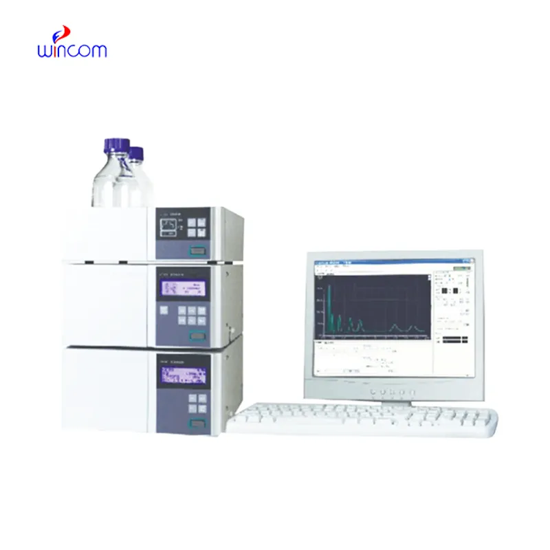
In hplc vial used to analyze metabolic profiles and biomarkers during clinical research laboratories. It enables the identification of disease markers and monitoring of biochemical changes over time through the separation of small molecules and proteins. hplc vial also facilitates the study of drug absorption and distribution, toxicity testing, and hospital-based clinical trials and thus making it possible to monitor patient responses to therapies in great detail while at the same time ensuring the accuracy and reliability of the analytical results.
hplc vial is assigned to become an important player in translational research which is being conducted in hospitals. Among the future developments are the combined detection systems, quicker analysis cycles, and improved reproducibility. hplc vial will be the mainstay of hospitals' molecular profiling and drug testing along with patient monitoring thus facilitating hospital diagnostics and personalized medicine research.
hplc vial proper care makes sure that hospital laboratories get reliable performance every time. Regular column flushing, taking care of pump and seal inspections, and using appropriate solvents are some of the measures that will keep the damage at bay and the separation efficiency high. Scheduled maintenance, system pressure control, and detector cleaning should be done by laboratory technicians to have longer instruments lifespan. Proper upkeep leads to less downtime and provides same quality analytical results in all clinical and research applications.
Clinical laboratories make use of hplc vial to analyze patient samples with remarkable accuracy. It identifies biomarkers, metabolites, and the levels of therapeutic drugs, thus giving reliable information about the disease status and monitoring treatment. Sensitivity of the technique permits determination of compounds in very minute amounts, which is critical in clinical testing. By resolving complex composition, hplc vial guarantees accurate and reproducible results for laboratory diagnostics. Lab staff utilizes it for daily testing, quality control, and research activities, thus making hplc vial a vital part of contemporary clinical laboratory work that caters to patient care, treatment choices, and lab data integrity.
Q: What is the sample preparation for HPLC? A: For the most part, samples should be filtered, diluted, or subjected to solvent extraction in order to avoid column clogs and have the results be accurate Q: Is HPLC able to pick trace-level compounds? A: With the right detectors, it can pick up such substances in extremely small amounts with high sensitivity. Q: Is HPLC a method that can be applied to analysis of proteins? A: Yes, particularly if one employs size-exclusion and reversed-phase columns for protein, peptide, and biomolecule separation. Q: What is the process of calibrating HPLC? A: The process is done by taking standards of known concentrations that are the same as the one in the sample and using them to check the performance of the column and the accuracy of the detector. Q: Are particular solvents needed for HPLC? A: Yes, the solvents used need to be compatible with the type of the column and the detectors to prevent any damage or interference in the analysis process.
The hospital bed is well-designed and very practical. Patients find it comfortable, and nurses appreciate how simple it is to operate.
This ultrasound scanner has truly improved our workflow. The image resolution and portability make it a great addition to our clinic.
To protect the privacy of our buyers, only public service email domains like Gmail, Yahoo, and MSN will be displayed. Additionally, only a limited portion of the inquiry content will be shown.
We’re interested in your delivery bed for our maternity department. Please send detailed specifica...
Could you share the specifications and price for your hospital bed models? We’re looking for adjus...
E-mail: [email protected]
Tel: +86-731-84176622
+86-731-84136655
Address: Rm.1507,Xinsancheng Plaza. No.58, Renmin Road(E),Changsha,Hunan,China

 af
af
 es
es
 ar
ar
 tr
tr
 sw
sw
 pt
pt
 th
th
 ur
ur
 bn
bn
 ne
ne
 vi
vi
 km
km
 lo
lo
 de
de
 ru
ru
 fi
fi
 nl
nl
 fa
fa
 fr
fr
 ko
ko