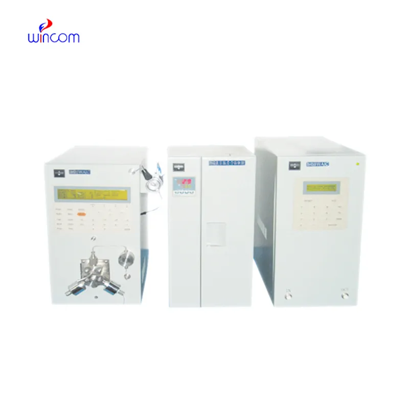
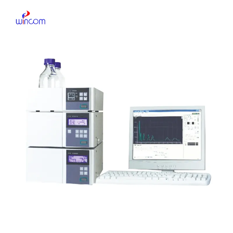
hplc instrumental offers high resolution separation of complex samples in clinical, pharmaceutical, and hospital laboratories, thereby supporting advanced laboratory workflows. It allows performing an in-depth analysis of drugs, metabolites, and small biomolecules. hplc instrumental is used by laboratory staff for research validation, patient monitoring, and method development. Its precision, speed, and adaptability make analytical efficiency greater and at the same time, make consistent and reproducible results which in turn, strengthen laboratory operations in the areas of healthcare and scientific environments.
hplc instrumental is indispensable in the hospital lab for vitamin and nutrient analyses of patient samples. It identifies and determines the amounts of vitamins and minerals that are deficient or excessive in blood or serum. Health care providers depend on it to keep track of patients' nutrition, provide aids for treatment, and assess the impact of supplementation which, thus, boosts the quality of clinical care overall and makes it more beneficial.
The instruments for hplc instrumental of the future will be equipped with separation methods in multiple dimensions and fully automated sample preparation. The detection of trace amounts of metabolites, drugs, and biomarkers will be so accurate that hospitals and clinical laboratories will be the first to reap the benefits. The applications of hplc instrumental in the future will greatly help in complex diagnostics, research studies, and laboratory efficiency.
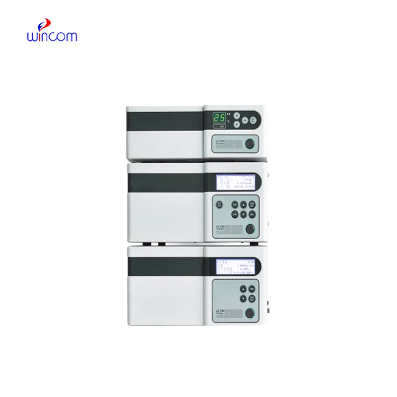
hplc instrumental proper care makes sure that hospital laboratories get reliable performance every time. Regular column flushing, taking care of pump and seal inspections, and using appropriate solvents are some of the measures that will keep the damage at bay and the separation efficiency high. Scheduled maintenance, system pressure control, and detector cleaning should be done by laboratory technicians to have longer instruments lifespan. Proper upkeep leads to less downtime and provides same quality analytical results in all clinical and research applications.
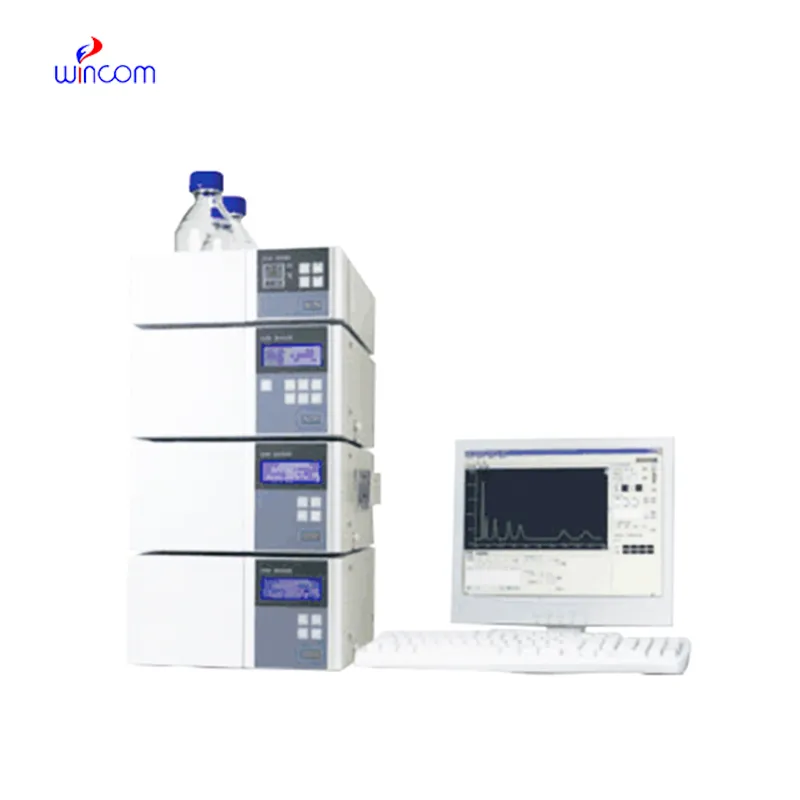
Therapeutic drug monitoring relies heavily on hplc instrumental in hospital settings. It determines the concentration of drugs in the body to guarantee efficiency and security. The laboratory staff uses it for the examination of blood, serum, or urine samples, and signifies small molecular compounds with high accuracy. By yielding consistent outcomes, hplc instrumental services the medics in changing the amounts and preventing side effects. Its use goes to hormone level testing, metabolite analysis, and pharmacokinetics research. With quick processing and accurate information, hplc instrumental is a part of the hospital patient care, making evidence-based treatment decisions possible and enhancing clinical outcomes in different departments.
Q: Do you need special training for HPLC operation? A: The answer is yes, training is a prerequisite to accurately and safely using pumps, columns, and detectors. Q: What type of maintenance does HPLC have? A: It requires cleaning, flushing, and inspection of all components as well as calibrating. Q: Is it possible to use HPLC in drug monitoring? A: Sure, it is a common practice in hospitals to monitor the levels of therapeutic drugs and also to identify metabolites in the samples taken from the patients. Q: What is the duration of analysis using HPLC in a typical case? A: The analysis time can range from a few minutes to more than an hour depending on the nature of the sample and the kind of column used. Q: Is HPLC a good choice for environmental testing? A: Yes, it can be used to find out the presence of pollutants, pesticides, and other harmful substances in water, soil, and air samples.
The microscope delivers incredibly sharp images and precise focusing. It’s perfect for both professional lab work and educational use.
I’ve used several microscopes before, but this one stands out for its sturdy design and smooth magnification control.
To protect the privacy of our buyers, only public service email domains like Gmail, Yahoo, and MSN will be displayed. Additionally, only a limited portion of the inquiry content will be shown.
Hello, I’m interested in your water bath for laboratory applications. Can you confirm the temperat...
We’re interested in your delivery bed for our maternity department. Please send detailed specifica...
E-mail: [email protected]
Tel: +86-731-84176622
+86-731-84136655
Address: Rm.1507,Xinsancheng Plaza. No.58, Renmin Road(E),Changsha,Hunan,China

 af
af
 es
es
 ar
ar
 tr
tr
 sw
sw
 pt
pt
 th
th
 ur
ur
 bn
bn
 ne
ne
 vi
vi
 km
km
 lo
lo
 de
de
 ru
ru
 fi
fi
 nl
nl
 fa
fa
 fr
fr
 ko
ko