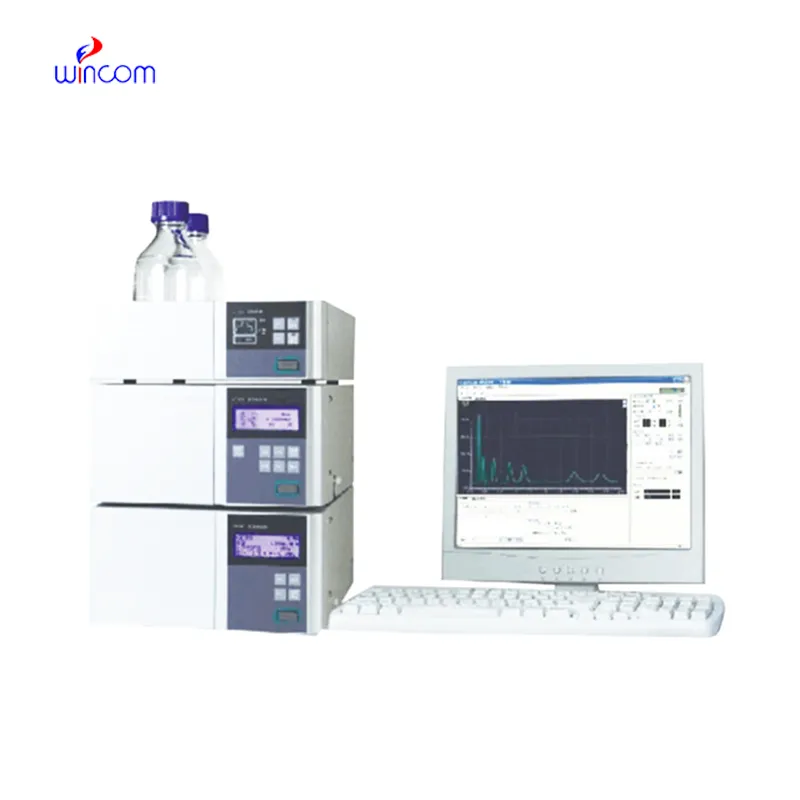
In the pharmaceutical lab, hplc analyse is the key to the precise assessment of the active substances, impurities, and metabolites. The machine gives a high-resolution separation, which in turn supports the quality assurance and the regulation compliance. Lab workers put their trust on hplc analyse for method validation, production consistency monitoring, and research trials. hplc analyse brings together the delicate ability to detect plus the repeated nature of results to make the complex formulations proficiently analyzed, thus, it serves the routine lab testing and the advanced experimental work in hospitals, research centers, and clinical facilities both.
Biochemical and clinical laboratories use hplc analyse to examine plasma or serum metabolites for disease research. It isolates and measures the amounts of small molecules participating in metabolism thus shedding light on patient conditions. The method is commonly employed in metabolic studies and experimental clinical trials conducted in hospitals.
The instruments for hplc analyse of the future will be equipped with separation methods in multiple dimensions and fully automated sample preparation. The detection of trace amounts of metabolites, drugs, and biomarkers will be so accurate that hospitals and clinical laboratories will be the first to reap the benefits. The applications of hplc analyse in the future will greatly help in complex diagnostics, research studies, and laboratory efficiency.
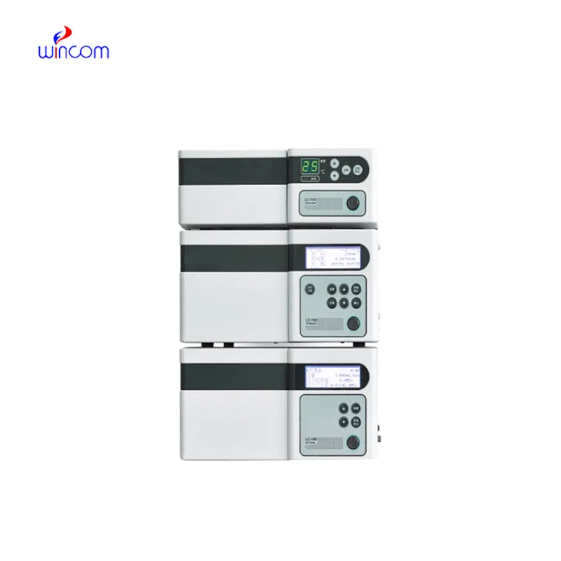
Proper handling and care of hplc analyse ensure continuous accuracy in the medical laboratory workflows. Cleaning of flow paths, checking detector response, and verifying pump performance are the essential maintenance tasks. Along with the column storage, solvent selection, and routine calibration, laboratory personnel must adhere to the manufacturer guidelines. Proper care enhances reproducibility, reduces downtime, and supports the consistent performance of the laboratory in hospitals and clinical research facilities.
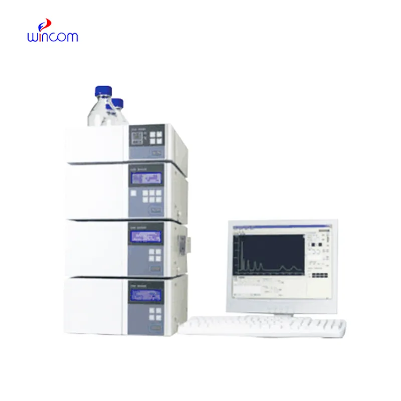
hplc analyse is a standard method in diagnostic laboratories of hospitals to keep an eye on patients’ biochemical and therapeutic figures. It quantifies drugs, hormones, and small molecules accurately. hplc analyse speeds up the clinical decision-making processes of physicians and facilitates treatment modifications by supplying them with quick and precise results. It is used by hospital labs for basic patient testing, pharmacokinetic studies, and special analyses. The method’s high reproducibility makes certain that the outcomes are consistent, whereas its versatility allows for the support of many clinical applications. hplc analyse has turned into an irreplaceable instrument in hospital diagnostics, which not only enhances patient management but also provides healthcare professionals with thorough molecular information.
Q: What is HPLC used for in laboratories? A: HPLC turns out to be one of the most significant and essential analytical methods in laboratories equipped with the chemical compound analysis, separation, identification, and quantification of their presence in complex samples which are the research, clinical, and pharmaceutical applications. Q: How does HPLC separate compounds? A: The HPLC separation technique is based on the different affinities of the compounds to the stationary phase and mobile phase within the chromatography column. Q: Can HPLC analyze biological samples? A: Yes, it is certainly possible to carry out analyses on various biological fluids such as blood, serum, urine, etc. for the detection of metabolites, drugs, and biomarkers. Q: How often should HPLC columns be replaced? A: The replacement of the columns must be done according to the manufacturer instructions or when the performance begins to decline, which is quite usual after heavy use or contamination. Q: What detectors can be used with HPLC? A: The analysis type determines the use of, among others, UV, fluorescence, refractive index, and mass spectrometry detectors as the common detectors.
We’ve been using this mri machine for several months, and the image clarity is excellent. It’s reliable and easy for our team to operate.
The hospital bed is well-designed and very practical. Patients find it comfortable, and nurses appreciate how simple it is to operate.
To protect the privacy of our buyers, only public service email domains like Gmail, Yahoo, and MSN will be displayed. Additionally, only a limited portion of the inquiry content will be shown.
I’m looking to purchase several microscopes for a research lab. Please let me know the price list ...
Hello, I’m interested in your centrifuge models for laboratory use. Could you please send me more ...
E-mail: [email protected]
Tel: +86-731-84176622
+86-731-84136655
Address: Rm.1507,Xinsancheng Plaza. No.58, Renmin Road(E),Changsha,Hunan,China

 af
af
 es
es
 ar
ar
 tr
tr
 sw
sw
 pt
pt
 th
th
 ur
ur
 bn
bn
 ne
ne
 vi
vi
 km
km
 lo
lo
 de
de
 ru
ru
 fi
fi
 nl
nl
 fa
fa
 fr
fr
 ko
ko