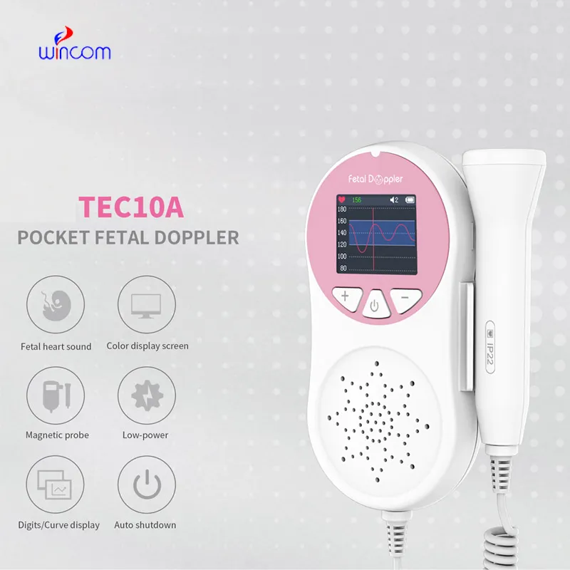
The fetal doppler at 13 weeks combines state-of-the-art digital imaging algorithms that boost contrast and depth perception, which leads to soft tissues being visualized more clearly. The intelligent interface features customizable scanning modes and user profiles for easier operation. Due to its low power consumption and sturdy construction, the fetal doppler at 13 weeks maintains performance that is even in high-demand clinical environments.

In medical imaging, the fetal doppler at 13 weeks is a trustworthy device for different clinical departments. It provides vascular studies support by observing the state of the arteries and veins; in urology, it has a role in the assessment of the bladder and prostate health. The fetal doppler at 13 weeks also enables emergency medical staff to make quick trauma evaluations, directing their actions precisely and efficiently.

The fetal doppler at 13 weeks can look forward to getting advantages from miniaturization and wearable technologies. Portable or handheld versions of the fetal doppler at 13 weeks will become more widespread to facilitate quick diagnoses in rural as well as emergency setups. The integration of telemedicine services will thus facilitate concurrent consultations via the fetal doppler at 13 weeks.

Care of the fetal doppler at 13 weeks involves much more than cleanup. Environmental monitoring and mechanical protection are also part of the process. The fetal doppler at 13 weeks should not be subject to either vibration or shock. The fetal doppler at 13 weeks should be backed up periodically to retain vital images.
Used in hospitals and clinics, the fetal doppler at 13 weeks provides immediate visual feedback for a variety of medical evaluation uses. Converting sound waves into live images, the fetal doppler at 13 weeks allows physicians to easily detect abnormalities. The fetal doppler at 13 weeks assists with making diagnostic processes safer in addition to improving patient outcomes. It possesses an ergonomic shape alongside digital integration capabilities that support simple data sharing and medical record documentation.
Q: What makes the ultrasound scannert effective for diagnostic imaging? A: Its high-frequency sound wave technology allows accurate visualization of internal body structures in real time. Q: How portable is the ultrasound scannert? A: The device features a compact and lightweight design, allowing easy movement between clinical departments. Q: What types of probes are compatible with the ultrasound scannert? A: It supports multiple probe types, including linear, convex, and phased array probes for varied diagnostic needs. Q: Does the ultrasound scannert require special training to operate? A: Basic technical training is recommended to maximize its imaging performance and functionality. Q: How long can the ultrasound scannert operate continuously? A: It is designed for extended use with efficient cooling systems and stable power performance.
The water bath performs consistently and maintains a stable temperature even during long experiments. It’s reliable and easy to operate.
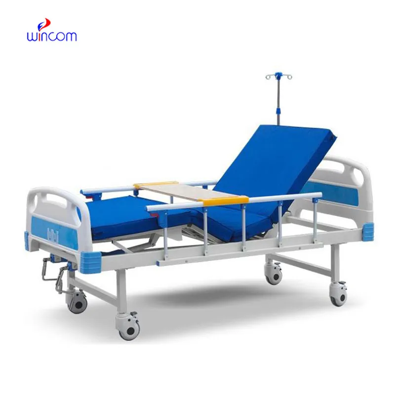
The delivery bed is well-designed and reliable. Our staff finds it simple to operate, and patients feel comfortable using it.
To protect the privacy of our buyers, only public service email domains like Gmail, Yahoo, and MSN will be displayed. Additionally, only a limited portion of the inquiry content will be shown.
We are planning to upgrade our imaging department and would like more information on your mri machin...
We’re interested in your delivery bed for our maternity department. Please send detailed specifica...
E-mail: [email protected]
Tel: +86-731-84176622
+86-731-84136655
Address: Rm.1507,Xinsancheng Plaza. No.58, Renmin Road(E),Changsha,Hunan,China

 af
af
 es
es
 ar
ar
 tr
tr
 sw
sw
 pt
pt
 th
th
 ur
ur
 bn
bn
 ne
ne
 vi
vi
 km
km
 lo
lo
 de
de
 ru
ru
 fi
fi
 nl
nl
 fa
fa
 fr
fr
 ko
ko