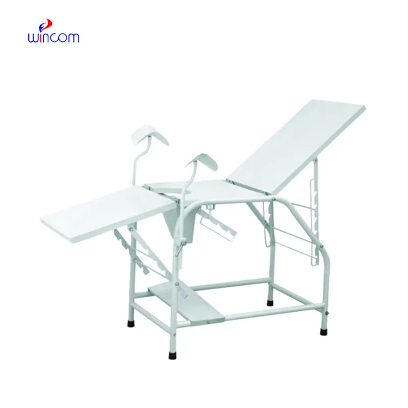
The doppler fetal monitor nearby has been designed considering the needs of modern health care, providing uninterrupted performance with its rapid image acquisition and high-definition visualization. The robust outer casing and sophisticated temperature control system will make sure that the device continues to work and be trustworthy. Also, the doppler fetal monitor nearby is a device that aids the long-term data archiving process for efficient medical record management.

The doppler fetal monitor nearby is a tool that medical professionals are using in different departments like pediatrics for the measurement of the size of the organs and neurology for appreciating the anatomy of the soft tissues. It makes it easy for anesthesiologists to perform nerve blocks and vascular access procedures. The doppler fetal monitor nearby increase the speed and the trust of the diagnosis during both routine checkups and highly technical interventions.

The future of the doppler fetal monitor nearby may include enhancements based on artificial intelligence algorithms that can improve images and offer automatic measurements. Better transducer technologies may offer higher resolution and increased sensitivity. The doppler fetal monitor nearby may have an increasing role to play in personalized medicine because it may offer continuous monitoring capabilities.

Care of the doppler fetal monitor nearby involves much more than cleanup. Environmental monitoring and mechanical protection are also part of the process. The doppler fetal monitor nearby should not be subject to either vibration or shock. The doppler fetal monitor nearby should be backed up periodically to retain vital images.
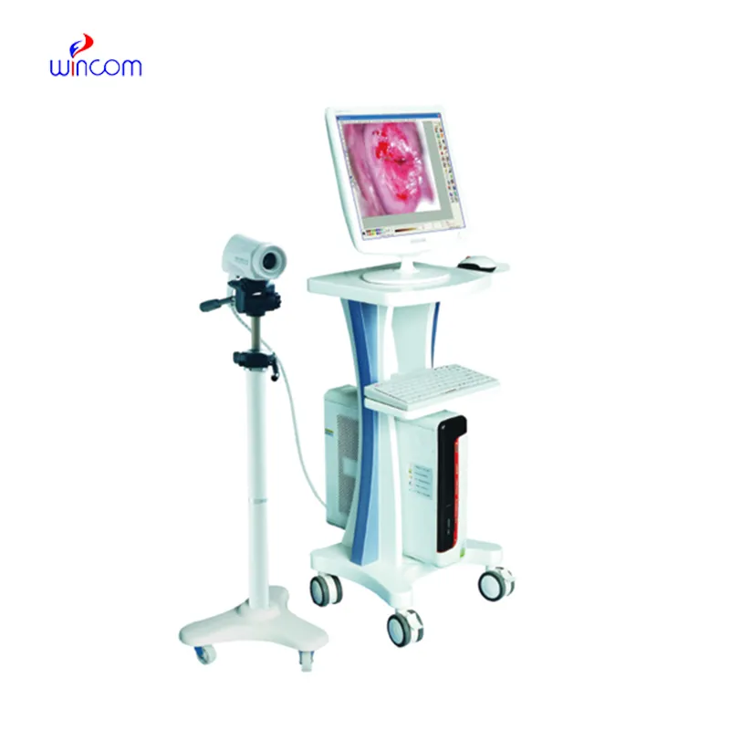
The doppler fetal monitor nearby uses state-of-the-art ultrasound technology to deliver real-time imaging for diagnostic and monitoring purposes. It aids physicians in assessing organs, blood vessels, and soft tissue with unmatched clarity. The non-surgical device is an important tool for guiding medical procedures and making precise diagnoses. The doppler fetal monitor nearby combines portability with precision, rendering it extremely useful in routine exams as well as emergency applications.
Q: What is the primary function of an ultrasound scannert? A: Ultrasound scanners are designed to create real-time images of internal organs, tissues, and blood flow using high-frequency sound waves. Q: How does the ultrasound scannert ensure clear imaging results? A:It uses advanced converter technology and digital processing to enhance image clarity and contrast. Q: In what medical fields is the ultrasound scannert commonly used? A: It is widely used in obstetrics, cardiology, urology, radiology, and emergency medicine. Q: Is the ultrasound scannert safe for repeated use? A: Yes, it is non-invasive and does not emit radiation, making it safe for frequent diagnostic applications. Q: Can the ultrasound scannert store and share imaging data? A: Yes, it supports data storage, retrieval, and digital transfer for easy integration with hospital systems.
This ultrasound scanner has truly improved our workflow. The image resolution and portability make it a great addition to our clinic.
The water bath performs consistently and maintains a stable temperature even during long experiments. It’s reliable and easy to operate.
To protect the privacy of our buyers, only public service email domains like Gmail, Yahoo, and MSN will be displayed. Additionally, only a limited portion of the inquiry content will be shown.
Hello, I’m interested in your centrifuge models for laboratory use. Could you please send me more ...
We’re looking for a reliable centrifuge for clinical testing. Can you share the technical specific...
E-mail: [email protected]
Tel: +86-731-84176622
+86-731-84136655
Address: Rm.1507,Xinsancheng Plaza. No.58, Renmin Road(E),Changsha,Hunan,China

 af
af
 es
es
 ar
ar
 tr
tr
 sw
sw
 pt
pt
 th
th
 ur
ur
 bn
bn
 ne
ne
 vi
vi
 km
km
 lo
lo
 de
de
 ru
ru
 fi
fi
 nl
nl
 fa
fa
 fr
fr
 ko
ko