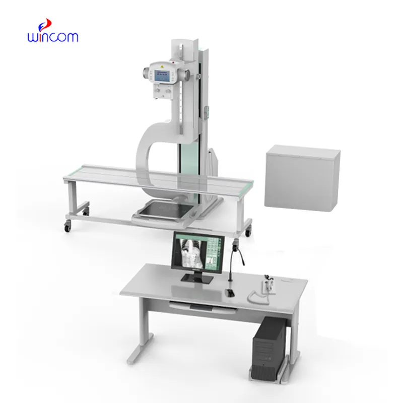
Designed with efficiency in mind, the diagram of an x ray machine combines exposure control and image processing. The equipment enables the observation of detail in both the bone and soft tissues without much distortion. The diagram of an x ray machine offers high flexibility when it comes to operations and can work well in various settings like hospitals and research labs.

The diagram of an x ray machine is commonly used in medical imaging to examine skeletal trauma, lung disease, and dental anatomy. The diagram of an x ray machine assists physicians in diagnosis of fractures, infection, and degenerative disease. The diagram of an x ray machine is also used in orthopedic surgery intraoperatively. In emergency medicine, it provides rapid diagnostic information that allows clinicians to assess trauma and internal injury rapidly.

Technological progress in the diagram of an x ray machine will provide faster image processing, improved 3D visualization, and more accurate diagnostics. Next-generation devices can have AI-assisted positioning systems which will preset imaging settings automatically. The diagram of an x ray machine will also be seamlessly integrated into cloud platforms in order to enable instant sharing of information as well as remote consultations.

The diagram of an x ray machine requires careful attention to ensure imaging accuracy and equipment safety. The housekeeping staff must test system performance on a regular basis, including tube voltage, exposure timing, and detector status. The diagram of an x ray machine should always be turned off when being cleaned and checked routinely for mechanical or electrical wear.
Owing to certain advances in modern technology, the diagram of an x ray machine that I’m writing about now uses digital radiography. Using digital radiography helps the diagram of an x ray machine offer improved diagnostic accuracy with less radiation exposure. The diagram of an x ray machine maintains supreme significance in diagnosing cases of fractures as well as joint and chest ailments.
Q: What is an x-ray machine used for? A: An x-ray machine is used to produce images of the internal structures of the body, helping doctors detect fractures, infections, and other medical conditions. Q: How does an x-ray machine work? A:X-ray machine emit controlled radiation that passes through the body and records varying degrees of absorption on detectors or film, creating visual images of bones and tissues. Q: Is it safe to use an x-ray machine frequently? A: Modern x-ray machines use very low doses of radiation, and protective measures such as lead aprons help minimize exposure for both patients and operators. Q: Can an x-ray machine detect soft tissue injuries? A: Although X-rays machine are primarily used to examine bones, they can reveal some soft tissue abnormalities, especially when used with contrast agents or digital image enhancement techniques. Q: Who operates an x-ray machine? A: X-ray machines are typically operated by trained radiologic technologists who ensure correct positioning, exposure settings, and safety protocols during imaging.
We’ve been using this mri machine for several months, and the image clarity is excellent. It’s reliable and easy for our team to operate.
The water bath performs consistently and maintains a stable temperature even during long experiments. It’s reliable and easy to operate.
To protect the privacy of our buyers, only public service email domains like Gmail, Yahoo, and MSN will be displayed. Additionally, only a limited portion of the inquiry content will be shown.
We are planning to upgrade our imaging department and would like more information on your mri machin...
I’d like to inquire about your x-ray machine models. Could you provide the technical datasheet, wa...
E-mail: [email protected]
Tel: +86-731-84176622
+86-731-84136655
Address: Rm.1507,Xinsancheng Plaza. No.58, Renmin Road(E),Changsha,Hunan,China

 af
af
 es
es
 ar
ar
 tr
tr
 sw
sw
 pt
pt
 th
th
 ur
ur
 bn
bn
 ne
ne
 vi
vi
 km
km
 lo
lo
 de
de
 ru
ru
 fi
fi
 nl
nl
 fa
fa
 fr
fr
 ko
ko